I. Introduction
The treatment of cancer has been receiving great attention from scientists for decades. The treatment of breast cancer has developed to a great extent in the past 20 years, and although the mortality rate of the disease has been greatly reduced, the incidence of the disease is still very high. Among the various types of breast cancer, "three-negative breast cancer" has been a difficult point of treatment because of the lack of therapeutic targets (such as estrogen receptor, progesterone receptor and human epidermal growth factor receptor 2). What is of interest to scientists is that "three-negative breast cancer" tumors have the most tumor-infiltrating lymphocytes, which are presumed to help fight tumor growth, whereas triple-negative breast cancer silences lymphocytes in tumors or Inactivated. This also gives us a hint that for triple-negative breast cancer, if we use immunotherapy to activate lymphocyte activity that kills tumors, it will be a new treatment strategy.
Second, what is the immunosuppressive mechanism?
The lecture first introduced the most important immunosuppressive mechanism, namely "PD-L1 and PD-1 mechanism of action" . Cancer cells express PD-L1 on the cell surface, and PD-L1 will link to PD1 of T cells. This association will eventually shut down the activity of T cells, that is, "immune escape" . Scientists have now developed monoclonal antibodies that specifically recognize PD-1 on PD-L1 or T cells on cancer cells. This strategy will help mask the surface of the interaction between the two molecules, thus blocking the linkage of PD-L1 and PD-1, thereby re-activating the activity of T cells, allowing T cells to recognize cancer cells, and then clearing these they. Currently, the overall response rate for this type of treatment is approximately 20%-40%. The speaker judged that there must be a similar immunosuppressive mechanism in "three-negative breast cancer."
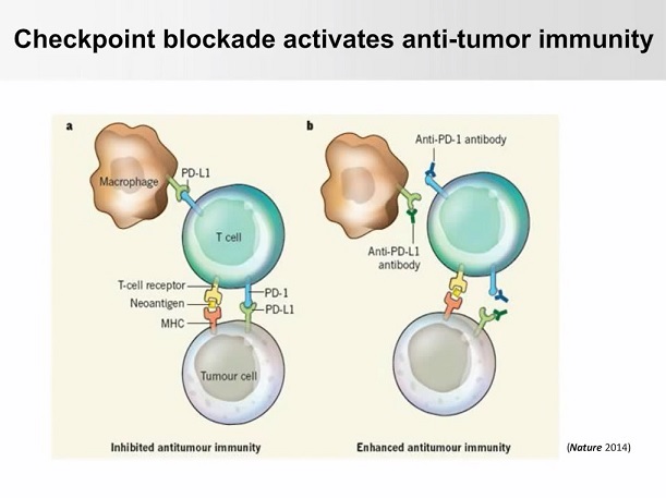
Figure 1. Mechanism of action of PD-L1 and PD-1
This study explored the mechanism by which monoclonal antibodies block "immune escape" and the development of new "antibodies and drug conjugates (ADC) by studying the glycosylation and stability of PD-L1 in triple-negative breast cancer cells. " To treat triple-negative breast cancer."
Third, experimental methods and results
The speaker first studied PD-L1 on cancer cells, trying to figure out which part of the cancer cells PD-L1 is expressed in? Since PD-L1 has a molecular weight of 33 kilodaltons, Western Blot experiments show that the protein expression of 33 kilodaltons is very small, while the protein of 50 kilodaltons is very abundant. After a series of gene knockout experiments, it was confirmed that this high molecular weight protein is the PD-L1 signal and is highly glycosylated PD-L1. It was found by mutation that PD-L1 only glycosylated on the four NXT domains in its extracellular region.
To validate the interaction of glycosylated PD-L1 and PD-1, the authors used the IncuCyte live cell dynamic imaging system to quantify dynamic changes between PD-L1 and PD-1. Green fluorescently labeled PD-1 protein was added to a solution of cancer cells (red nuclei) with glycosylated PD-L1. At 12 hours after treatment, there was a large amount of green aggregation on the cell surface, indicating PD-L1 and PD-1 has an interaction. For the stable clone of non-glycosylated PD-L1, no green fluorescence was observed, which means that PD-L1 and PD-1 did not interact after 12 hours. The results show that this glycosylation structure is required for protein interactions.

Figure 2. Quantification of dynamic changes between PD-L1 and PD-1 using an IncuCyte imager
To understand the function of glycosylation, the speaker again used the IncuCyte imager to simulate the dynamic interaction of cancer cells and T cells in the presence of T cells. In this example, for BT549 nuclear IP cells, activated T cells were added and incubated for 5 days, and apoptosis of cancer cells was expressed by green fluorescence indicating Caspase 3 activity. As long as the cancer cells begin to die, you can see a decrease in red fluorescence and an increase in green fluorescence. The results showed that cancer cells with glycosylated PD-L1 were resistant to T cell killing, while non-glycosylated cancer cells were very sensitive to T cell killing.
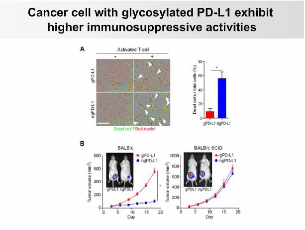
Figure 3. IncuCyte imager to simulate the dynamic interaction of cancer cells and T cells in the presence of T cells
Next, the speaker wants to know what is the upstream signal that regulates glycosylation? They treated two triple-negative breast cancer cells, BT549 and BT468, with different stimuli such as EGF, IGF, HGF, FGF and TGFβ. EGF was found to consistently induce glycosylation of PD-L1 in two triple-negative breast cancer cells. The glycosylated PD-L1 is very stable, while the non-glycosylated PD-L1 has a short half-life. The upstream signal EGF can increase the glycosylation pattern of PD-L1. So if we inhibit EGFR signaling, we can make triple-negative breast cancer cells in a homologous animal model sensitive to the treatment of "anti-PD-1".
Another finding is that triple negative also induces chronic inflammation. They want to understand why these inflammations can cause cancer to develop. The speaker found that in the tumor microenvironment, macrophages have the ability to secrete TNFα (tumor necrosis factor alpha), while TNFα recognizes the TNFα receptor of cancer cells. TNFα will activate NF-kappaB and cause nuclear transfer of p65, which then initiates expression of CSN5. CSN5 is a ubiquitinating enzyme that will stabilize PD-L1, induce immunosuppressive function of cancer cells, and promote the development of cancer cells.
The speaker collaborated with the pharmaceutical company to develop antibodies against glycosylation of PD-L1. They inoculated glycosylated PD-L1 as an antigen on mice, creating 3,000 hybridoma clones. These 3,000 clones were then screened by flow cytometry in an effort to find antibodies that specifically recognize glycosylated PD-L1 without identifying non-glycosylated PD-L1. The IncuCyte imager was then used to further reveal which antibody has the function of blocking the PD-L1 and PD-1 interactions. They used Essen BioScience's technology and the pHrodo phagocytic system to study whether antibodies bind to glycosylated PD-L1, then induce the internalization of this PD-L1 and enter the lysosomes to be degraded by cells. The speaker labeled the STM108 antibody with a fluorescent dye for fluorescence. When the cells begin to phagocytose these antibodies, red fluorescence will be observed in the IncuCyte imager . In this case, when they added the STM108 antibody to a stable clone of PT549, it was observed that a very beautiful red fluorescence was induced during the 12-hour period, suggesting that the STM108 antibody can be specifically internalized into cancer cells. . Then Western Blot was done after 5 days of treatment, and a large amount of PD-L1 was found to be reduced, suggesting that PD-L1 was degraded .
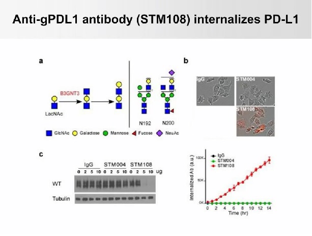
Figure 4. Observation of the pHrodo phagocytic system with an IncuCyte imager, suggesting that the STM108 antibody can be specifically internalized into cancer cells.
The speaker also studied the antibody and drug binding technology, selected MMAE as a warhead, and bound cytotoxic drugs to the antibody. When mice had PD-L1 expression, their tumor growth was significantly reduced after treatment with "antibody drug conjugates (ADC) . 4T1-hPDL1 tumors no longer grew after treatment with glycosylated ADC antibodies. When the mice were treated with glycosylated MMAE antibodies, strong red fluorescence was observed in the tumor microenvironment, indicating an increase in the activity of Caspase 3 and apoptosis in the cells. Once an endocytic event occurs, PD-L1 protein will enter the lysosome and be degraded. With the degradation of antibodies and PD-L1, cytotoxic drugs against cancer cells will be released, and then the cancer with high expression of PD-L1 will be killed. cell. This also produces a "bystander effect" that removes cancer cells that do not express PD-L1.
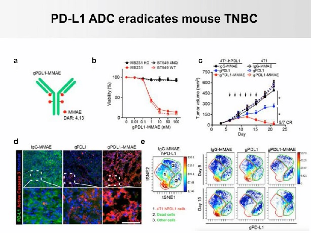
Figure 5. Observing red fluorescence in the tumor microenvironment, indicating increased activity of Caspase 3
Fourth, the conclusion
Based on their signal analysis, the speaker can design strategies to isolate monoclonal antibodies that specifically recognize glycosylated PD-L1. They first discovered that this anti-PD-L1 antibody can cross-link the "PD-L1" molecule bound to the membrane and induce endocytosis of this protein, thus allowing the antibody to carry cytotoxic drugs and release the drug by endocytosis. Kill cancer cells and trigger a profound effect on triple-negative breast cancer.
V. Automated live cell dynamic analysis
At present, most of the cell detection methods still belong to the traditional end point method - only the final result is given. This method can not only dynamically monitor and analyze the whole process of cell experiments, but also obtain biased results. In order to be able to observe live cells throughout the process, Essen Corporation of the United States has developed the IncuCyte S3 "Viable Cell Dynamic Imaging and Analysis System" built into the incubator, inside the incubator, and simultaneously for 6 cell culture dishes/bottles/microplates (6 -384 wells) for long-term dynamic imaging, recording real-time changes in cell morphology details, and transforming into a variety of cell function-related time-quantitative curves through powerful image analysis software.
IncuCyte S3 is a cell-level scientific and drug screening solution for immunosuppression, nerve growth and regression, cancer cell metastasis, cell migration, stem cell differentiation, apoptosis, cell necrosis, cell proliferation, wound repair, etc. Large-scale screening and research on long-term life science issues.
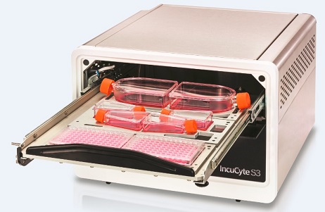
Figure 6. IncuCyte S3 Live Cell Dynamic Imaging and Analysis System
Sixth, online lecture video
Video duration is 1hr8min, please watch under WiFi conditions:
Https://v.qq.com/x/page/n05302ljxpr.html
7. About "Diaoao Biotechnology"
“ Diao Biotech †(Shanghai, Hong Kong, Taiwan) was established in 1994. For more than 20 years, it has been adhering to the world-class instrument manufacturers. The experts with rich R&D experience are technical support and the professional maintenance team serves. Backed by a focus on providing state-of-the-art hardware and software equipment and technical services for life sciences, pharmaceutical R&D and disease treatment.
The exclusive distributors of " Diao Biotech " are:
IncuCyte, Beacon, Chipcytometry, Gyrolab, iQue Screener PLUS, Avatar, PlexBio, Certus, HighRes, etc.
Telephone (transfer to the marketing department)
Email:
Website:
Scan the QR code to add the WeChat public account " tekontech " :

Fruits And Vegetables,Fresh Fruits And Vegetables,Fresh Fruit And Veggies,Fresh Vegetables
Xi'an Gawen Biotechnology Co., Ltd , https://www.seoagolyn.com