In recent years, the ability to improve plant function and structure with high precision through non-destructive testing has become the main goal of plant breeding and precision agriculture. The emerging research methods of plant phenotype reveal plant growth, yield, quality and resistance to various stresses. Quantitative traits play a key role. In addition to the fully automated phenotypic analysis system, other cost-acceptable high-throughput protocols can help researchers more accurately grasp various plant traits, such as hyperspectral imaging and chlorophyll fluorescence imaging (Pasquale Tripodi et al., 2018). year).
[Case 1] Using hyperspectral imaging and chlorophyll fluorescence imaging technology to detect the perishable degree of fresh-cut lettuce
The American Agricultural Research Institute and the Australian High Resolution Plant Phenotypic Center have jointly studied the perishable levels of nine lettuces in the market's common Modified atmosphere packaging (MAP) (dissertation published in Postharvest Biology and Technology) , 2015). Because lettuce is highly perishable and difficult to detect by visual inspection in the early stages of decay, a system for early detection and assessment of changes in lettuce production and breeding quality for lettuce processing and breeding companies is available. It is especially important.
For the convenience of reading and understanding, the editor lists the names and physical photos of the nine varieties of lettuce, see below. 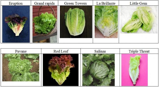 Â The researchers developed two lettuce decay index (LEDI), which are index LEDI 4 based on three-band hyperspectral imaging and index LEDI CF based on chlorophyll fluorescence imaging. In addition to detecting lettuce rot, the index can also Identifying tissues damaged by low temperature freezing, for red, dark green, green, light green, and yellow leaves, the two indices achieve nearly 97% accuracy in fresh/decay grading.
 The researchers developed two lettuce decay index (LEDI), which are index LEDI 4 based on three-band hyperspectral imaging and index LEDI CF based on chlorophyll fluorescence imaging. In addition to detecting lettuce rot, the index can also Identifying tissues damaged by low temperature freezing, for red, dark green, green, light green, and yellow leaves, the two indices achieve nearly 97% accuracy in fresh/decay grading. 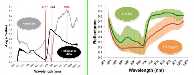 A graphical representation of the difference in reflectivity between fresh and decayed tissue, based on hyperspectral imaging data from 100 fresh and decayed samples
 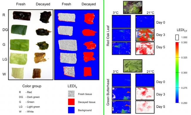 The left picture shows the fresh/decided visual grading of 5 groups of lettuce samples and the hyperspectral grading based on LEDI 4 index. The figure on the right shows the chlorophyll fluorescence imaging and corresponding LEDI CF index of two kinds of lettuce after different temperature treatment.
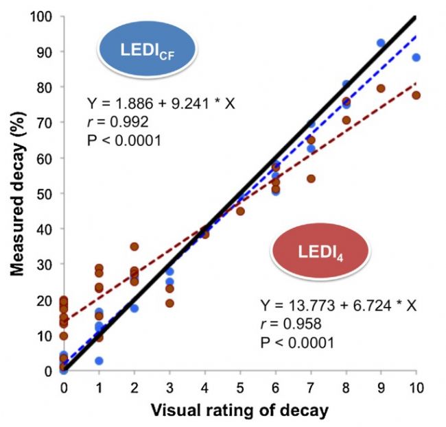
[Case 2] Chlorophyll fluorescence and hyperspectral imaging nondestructive detection of glyphosate-resistant transgenic corn shikimic acid concentration
Researchers at Zhejiang University and the Academy of Agricultural Sciences have used visible-near-infrared hyperspectral imaging and chlorophyll fluorescence imaging techniques to combat glyphosate-type transgenic maize (Frontiers in Plant Science, 2018), for both wild-type and transgenic. The water-spraying and glyphosate spraying experiments were carried out in the three-leaf stage maize seedlings, and the shikimic acid concentration in the leaves of the plants was determined by chemical methods. The results showed that the partial least squares regression model established at the optimal wavelength effectively predicted. The determination coefficients of shikimic acid concentration, calibration group and prediction group reached 0.79 and 0.82, respectively. Moreover, predicting shikimic acid concentration by visualizing spectral images helped to develop a simple multi-spectral imaging instrument for non-destructive phenotypic detection, and a new data method was combined. Chlorophyll fluorescence imaging also provides a satisfactory fit model for shikimic acid concentration. 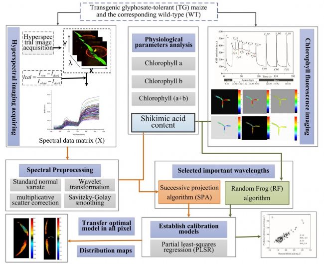
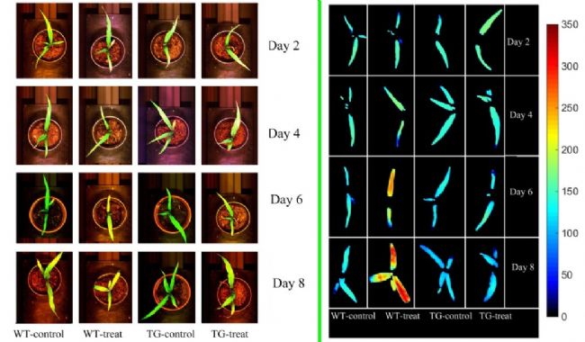
 The chlorophyll fluorescence imaging of the PSII maximum photon yield (F v /F m ) of the entire corn canopy during glyphosate treatment, the color bars on the right indicate the range of values ​​and the relationship to the color map
Yi Ketai provides you with the above non-destructive testing technology solutions
Specim hyperspectral imaging technology
Hyperspectral imaging is a new crop detection technology that combines traditional imaging and spectroscopy to obtain both spatial and spectral information for a sample. Due to its powerful capabilities, it has been widely used in crop nutrient detection, disease diagnosis and growth status monitoring. Specim hyperspectral imaging technology can not only detect plant health or disease, but also identify the types of plant diseases, grade the severity, and judge the disease period. 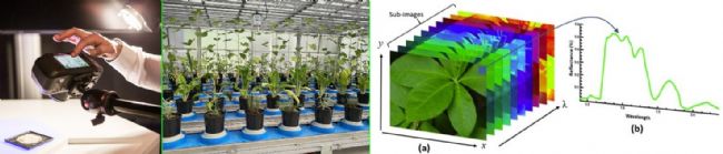
FluorCam multispectral fluorescence imaging
FluorCam multispectral fluorescence imaging technology is based on FluorCam chlorophyll fluorescence imaging technology, which can be used for both chlorophyll fluorescence dynamic imaging analysis and long-wavelength UV ultraviolet light (320nm -400nm) for multi-spectral fluorescence imaging of plant leaf excitation. Measurement analysis. FluorCam chlorophyll fluorescence imaging has long been an important instrument for detecting plant biotic and abiotic stresses. FluorCam multispectral fluorescence imaging can detect primary and secondary metabolites produced by plants and is widely used in plant disease detection, especially in early plant disease detection. 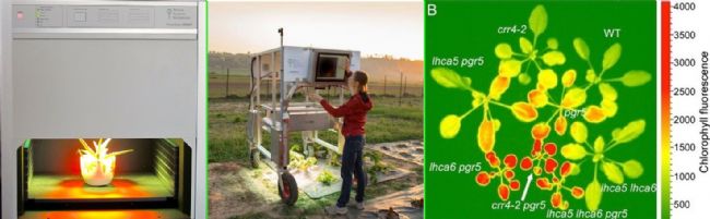
Thermo-RGB infrared thermal imaging technology
Infrared thermal imaging technology has the advantages of high sensitivity, non-contact, high image precision, wide measurement range and easy to realize automatic real-time observation. It is a hotspot of non-destructive testing research. Thermo-RGB infrared thermal imaging technology combines infrared thermal imaging technology with RGB technology to not only detect RGB images of diseased plants, but also detect differences in plant surface temperature during early infection of diseases by infrared thermography, revealing stomatal heterogeneity after disease stress. Sexual opening and closing, reflecting the early characteristics of crop infections. 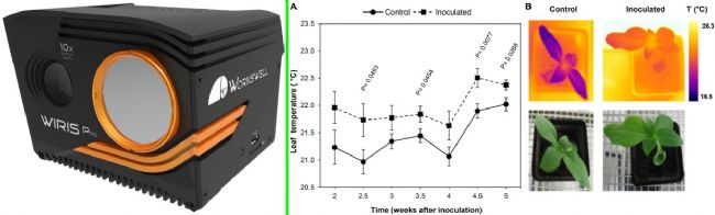
[Case 1] Using hyperspectral imaging and chlorophyll fluorescence imaging technology to detect the perishable degree of fresh-cut lettuce
The American Agricultural Research Institute and the Australian High Resolution Plant Phenotypic Center have jointly studied the perishable levels of nine lettuces in the market's common Modified atmosphere packaging (MAP) (dissertation published in Postharvest Biology and Technology) , 2015). Because lettuce is highly perishable and difficult to detect by visual inspection in the early stages of decay, a system for early detection and assessment of changes in lettuce production and breeding quality for lettuce processing and breeding companies is available. It is especially important.
For the convenience of reading and understanding, the editor lists the names and physical photos of the nine varieties of lettuce, see below.
 Correlation diagram of chlorophyll fluorescence index LEDICF and hyperspectral index LEDI4
[Case 2] Chlorophyll fluorescence and hyperspectral imaging nondestructive detection of glyphosate-resistant transgenic corn shikimic acid concentration
Researchers at Zhejiang University and the Academy of Agricultural Sciences have used visible-near-infrared hyperspectral imaging and chlorophyll fluorescence imaging techniques to combat glyphosate-type transgenic maize (Frontiers in Plant Science, 2018), for both wild-type and transgenic. The water-spraying and glyphosate spraying experiments were carried out in the three-leaf stage maize seedlings, and the shikimic acid concentration in the leaves of the plants was determined by chemical methods. The results showed that the partial least squares regression model established at the optimal wavelength effectively predicted. The determination coefficients of shikimic acid concentration, calibration group and prediction group reached 0.79 and 0.82, respectively. Moreover, predicting shikimic acid concentration by visualizing spectral images helped to develop a simple multi-spectral imaging instrument for non-destructive phenotypic detection, and a new data method was combined. Chlorophyll fluorescence imaging also provides a satisfactory fit model for shikimic acid concentration.
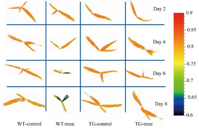
Determination of shikimic acid process in corn leaves based on chlorophyll fluorescence imaging and hyperspectral imaging
The left picture shows the change of corn leaf with time under visible light; the upper part of the figure shows the visual classification by high spectrum, and the color bar from blue to red indicates the increase of shikimic acid concentration.
Note: WT stands for wild-type, TG stands for transgenic, control is sprayed with water, and treat is sprayed with glyphosate. Yi Ketai provides you with the above non-destructive testing technology solutions
Specim hyperspectral imaging technology
Hyperspectral imaging is a new crop detection technology that combines traditional imaging and spectroscopy to obtain both spatial and spectral information for a sample. Due to its powerful capabilities, it has been widely used in crop nutrient detection, disease diagnosis and growth status monitoring. Specim hyperspectral imaging technology can not only detect plant health or disease, but also identify the types of plant diseases, grade the severity, and judge the disease period.
FluorCam multispectral fluorescence imaging
FluorCam multispectral fluorescence imaging technology is based on FluorCam chlorophyll fluorescence imaging technology, which can be used for both chlorophyll fluorescence dynamic imaging analysis and long-wavelength UV ultraviolet light (320nm -400nm) for multi-spectral fluorescence imaging of plant leaf excitation. Measurement analysis. FluorCam chlorophyll fluorescence imaging has long been an important instrument for detecting plant biotic and abiotic stresses. FluorCam multispectral fluorescence imaging can detect primary and secondary metabolites produced by plants and is widely used in plant disease detection, especially in early plant disease detection.
Thermo-RGB infrared thermal imaging technology
Infrared thermal imaging technology has the advantages of high sensitivity, non-contact, high image precision, wide measurement range and easy to realize automatic real-time observation. It is a hotspot of non-destructive testing research. Thermo-RGB infrared thermal imaging technology combines infrared thermal imaging technology with RGB technology to not only detect RGB images of diseased plants, but also detect differences in plant surface temperature during early infection of diseases by infrared thermography, revealing stomatal heterogeneity after disease stress. Sexual opening and closing, reflecting the early characteristics of crop infections.
The slim Casual Pants is suitable for men's daily travel. The pants have a few simple bags, which sre convenient for you to carry things out. The fashionable wide - brimmed waistband design is foiled the strong and muscular figure of men. The paste cloth back bag is simple fashion generous. Our factory can take long-term FOB orders, more than 100 pieces can do goods.Welcome to tae sample purchase.
100% Cotton Casual Pants,Cotton Baggy Trousers,Cotton Linen Loose Pants,Casual Cotton Blend Pants
Xinhui Jielide Garment Factory , https://www.ntgfwear.com